A pregnancy test detected the fetus in the test tube. The artificial models developed from stem cells - without egg, sperm or uterus - and grew in the laboratory until day 14.
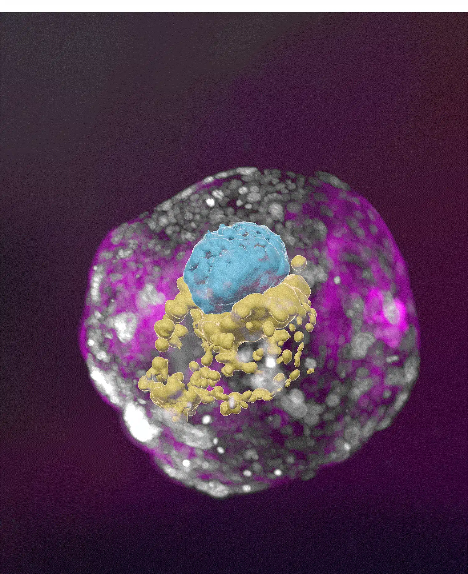
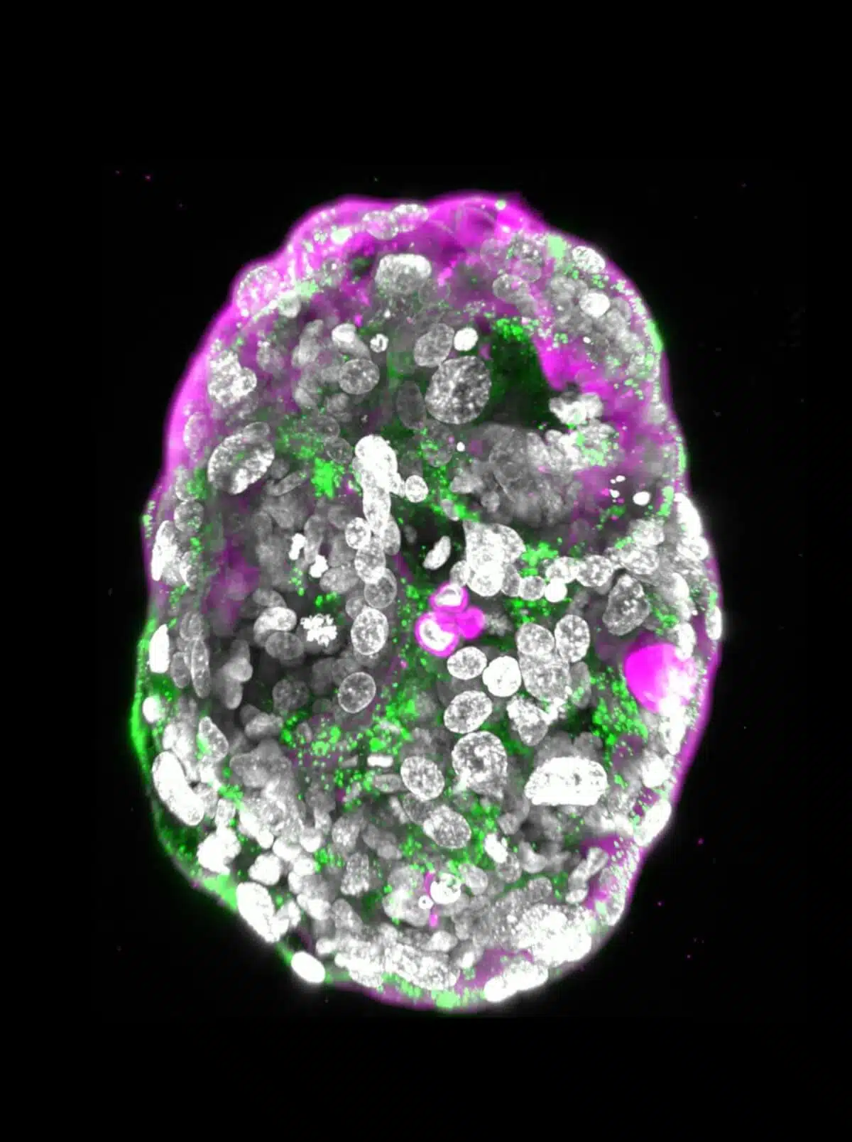
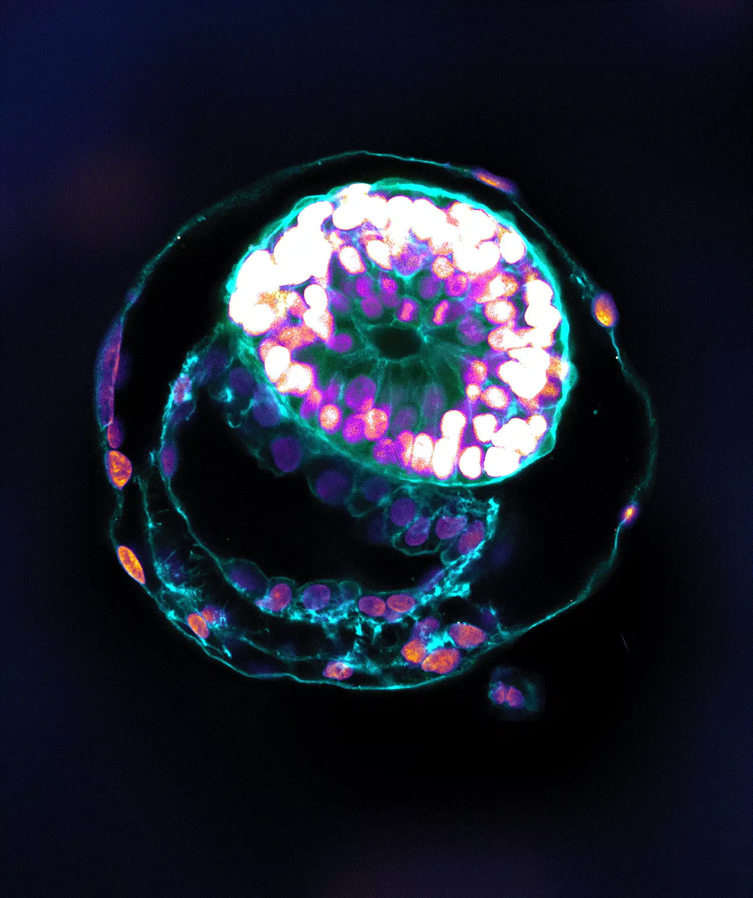
What differentiates between a human embryo in its early stages and a cluster of cells developing in a laboratory dish? Already from day 7 onwards, the accumulation of cells that will become a person produces an orderly and wonderful structure - a true master plan that dictates the manner of its development in the following months. In a new study published today in the scientific journal Nature, the scientists of the Weizmann Institute of Science managed to reproduce this unique structure and created artificial models of human embryos. The team of researchers from the laboratory of Prof. Yaacov Hana in the department of molecular genetics at the institute created the artificial models from human stem cells and grew them outside the womb until day 14. The human models they created are faithful to the original, meaning they contain all the structural components and systems characteristic of this stage in the fetus, including the placenta, The yolk sac, the amniotic sac and the other external tissues necessary for normal development.
In previous studies, clusters of cells developed from human stem cells were indeed created, but these were not recognized as faithful models of the origin of human embryos because they lacked almost all the key characteristics of an embryo. For example, they did not include some of the essential cell types for the developing embryo, including the outer layer of cells of the fertilized egg that develops into the placenta and the amniotic sac. In addition, they were not endowed with the unique structural organization that characterizes the embryo and did not show dynamic signs indicating the ability to advance to the next stage of development.
Reliable models of the beginning of life have far-reaching significance in the worlds of science and medicine, as very little is currently known about the earliest stages of the human embryo. Thanks to their close proximity to the embryonic source, the new models will provide an unprecedented glimpse into the beginning of embryonic development and dispel the shroud of mystery surrounding this primordial process. "The drama of human embryonic development takes place in the first month - the remaining eight months are largely the continuation of the course outlined in the first month," says Prof. Hana. "However, the first month is still largely a black box, both for ethical reasons that limit the ability to study these processes in human embryos and for technical difficulties. Our artificial model offers an ethical and accessible way to peer into this box; It reliably imitates the development of the natural embryo and the formation of its complex and wonderful structure."
When the model roars "Come on!"
When it came to creating models of human embryos, Prof. Hana's research team was based on previous breakthroughs of the laboratory and especially on the research published last year in the scientific journal Cell and presented for the first time an artificial model of mouse embryos from stem cells only. Similar to the mouse model, the human model does not use a uterus, egg or sperm, but only stem cells that have been returned to the so-called "naive" state - the earliest developmental stage in which the stem cells have the ability to differentiate into all types of cells and tissues; This situation corresponds to day 7 of a natural human embryo - the rooting stage in the uterine wall. The cells used by the researchers were mature skin cells that were returned to the state of stem cells using a method developed in Prof. Hana's laboratory, as well as stem cells that had been cultured in the laboratory for years.
The scientists divided the stem cells into three groups. The first group, from which the embryo itself developed, remained unchanged, while the other two groups were treated with chemicals, with the aim of increasing the expression of genes necessary for the creation of three types of extra-embryonic tissues that support the development of the embryo - the placenta, the yolk sac and the amniotic sac. Later, cells from the three groups were mixed under unique conditions developed in the laboratory, and began to develop into clusters of cells. However, only about 1% of the cell clusters eventually developed into embryo-like structures in a spontaneous process of self-assembly. Prof. Hana explains: "By its very essence, the fetus wants to develop. We don't need to tell him what to do, but only to release the potential encoded in him. But for this to happen, the right cells need to be mixed - naive stem cells that are not limited in terms of differentiation. Once these conditions are met, the embryo-like model roars 'Go!'"
The pregnancy test came back positive
The clusters of cells formed from the stem cells developed outside the womb for 8 days and reached the stage corresponding to day 14 in the development of the human embryo; The point in time when the natural embryo moves to the next developmental stage - the creation of the tissues from which the various organs of the body will develop. When the scientists compared the models they created with diagrams and microscope images of human embryos as published in classic textbooks from the sixties of the last century, they discovered a clear resemblance to the natural embryos. Every structural component and every supporting tissue was found in the models in their intended place, and were normal in terms of shape and size. Even the cells that secrete the hormone used in pregnancy tests were in the right place, and they also worked properly: when the scientists dripped these secretions onto a commercial pregnancy test kit, it returned a positive answer.
The researchers' new approach is based on many years of research in Prof. Hana's group, which was also the first to isolate stem cells in a naïve state - an essential "building block" for the success of an authentic embryonic model. This approach is expected to give rise to many research directions: for example, it may help decipher the causes of various birth defects and fertility problems; Lead the development of new technologies for growing tissues and organs for transplantation - and offer an alternative to experiments that cannot be performed on human embryos, such as testing the effect of drugs or other substances on fetal development.
In fact, already during their current work, the scientists discovered a finding that may shed light on early abortions, including those that occur when the woman does not even know she is pregnant. They saw that the cells from which the placenta develops must be organized correctly in space already on the third day of the development of the artificial models - day 10 in natural embryos - and that their failure to appear in a normal way means abnormal fetal development. Prof. Hana says: "Many abortions occur at the beginning of pregnancy. Many congenital defects are also created in these stages, which will be discovered in much later stages. Our models are expected to make it possible to reveal the biochemical and mechanical signals necessary for normal fetal development at this early stage, and also reveal the disruptions that may happen during the normal course of affairs."
Dr. Bernardo Oldak, Emily Wildschutz, Dr. Vladislav Bondarenko, Alejandro Aguilera, Shadi Terzi, Mamet Yunus Komar, Shahed Ashiohi, Dmitry Lokshtanov, Dr. Francesco Roncto, Sergey Vyukov, Eitan Ariel, Max Rose, participated in the study. Nir Livnat, Dr. Thom Shani, Karin Jobran, Roni Cohen and Dr. Noa Noverstern from Prof. Hana's laboratory in the department of molecular genetics of the institute; Dr. Yosef Addi and Dr. Merav Kadami from the Department of Life Science Research Infrastructures of the Institute; Dr. Hadas Keren Shaul from the Israeli National Center for Personalized Medicine named after Nancy and Steven Grand; Dr. Cheng Zhao, Prof. Sophie Petropoulos and Prof. Frederik Lanner from the Karolinska Institute in Sweden; and Prof. Vincent Pask from the University of Leuven in Belgium.
More of the topic in Hayadan:

6 תגובות
Why only 14 days? What happens after 14 days? Come and hear my lecture on the subject
I don't know if he can't, but it's definitely not allowed by law.
It seems that an embryo from stem cells cannot be implanted in a human womb and become a live embryo. Not yet. I would love to be fooled.
There is no difference except that it is not implanted in the womb and it does not become a person.
Can you shed light on the difference between a "model of an embryo" and an embryo?
Can you shed light on the difference between a model of an embryo and a fetus?