Artificial respiration is a life-saving technology, but it can damage the respiratory system. Researchers at the Technion present new aspects of this injury and offer preliminary treatment that will moderate it among breathing premature infants and thus improve their health
About a tenth of the babies in the world are born prematurely, before their body systems had time to develop properly. One of these systems is the respiratory system, which reaches full maturity at the age of two and is essential for the baby's survival. When the fetus is in the womb, it receives oxygen from its mother through the umbilical cord, but after birth it must breathe on its own. Therefore, premature babies are often characterized by respiratory distress and need support to breathe. Furthermore, the earlier the premature baby is born, the more severe the respiratory distress is and he is expected to have a longer respiratory period. Sometimes, the ventilation is done using a ventilator that puts air into the lungs through a tube inserted into the trachea.
Despite the continued improvement in respiratory technologies and treatment of premature babies, the morbidity rates among them are still higher than desired. One of the problems stems from the fact that essential treatments in prematurity, including artificial respiration, do indeed prevent death but create damage and side effects that may affect the patient in the long term. These effects and sources of harm from artificial respiration are not yet fully understood. This is the background for the research done in the laboratory of Prof. Joshua Schnittman from the Faculty of Biomedical Engineering at the Technion.
In a previous study, published last year inJournal of the Royal Society Interface, Prof. Joshua Schnittman and Dr. Eliram Nof discovered a phenomenon that is not mentioned at all in the medical literature: lung damage as a result of an air jet exiting the tube inserted into the trachea of the premature infant during artificial respiration. Using a physical model (flow mechanics) the two discovered that this jet exerts strong shearing forces on the epithelial tissue - the layer of cells that lines the airways. These forces can cause damage that endangers the patient, and even more so when it comes to a premature baby.
In the follow-up study, now published in the journal Bioengineering & Translational Medicine, the researchers tested the hypothesis in a new model containing an artificial human epithelium. The research was led by Prof. Schnittman, Dr. Eliram Nof and Dr. Arbel Artzi-Schnirman. Pediatricians and otolaryngologists are also involved in the research, including Dr. Liron Bornstein, a faculty member at the Rappaport Faculty of Medicine at the Technion and a senior physician in the Neonatal and Premature Intensive Care Department at the Rambam Medical College.
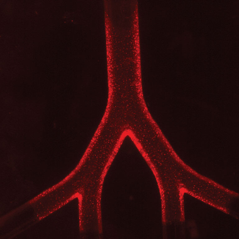
The research was done using a XNUMXD model of the upper airways - the trachea and the bronchi that diverge from it. Within this model, the researchers grew a layer of epithelial cells - the cells that also cover the natural respiratory system - to monitor the effect of breathing on them. "Today we already know that artificial respiration, which is a life-saving technology, causes various damages to the respiratory system," explains Prof. Schnittman. "These damages have so far been attributed to mechanical factors such as the entry of pressurized air that causes the lung tissue to stretch. In recent years, insights into more complex sources of damage have been added. In the current study, we demonstrated in vitro experiments by measuring cytokines that activate the immune system, the initiation of the inflammatory process - a process that is at the root of the lung damage in ventilated premature infants resulting from the air flow and shear forces during respiration."
In the previous study, the Technion researchers discovered, as mentioned, that the air jet exerts strong shearing forces on the epithelial tissue; Now they have discovered that the jet causes a change in the balance of cytokines - proteins that form the basis of communication between immune system cells. In other words, beyond the mechanical damage - the erosion of the epithelium - the invasive ventilation also causes an immune response to be activated and this may lead to harmful and even life-threatening inflammatory developments.
Following these discoveries, the researchers examined the possibility of reducing the said damage through drug treatment, and with the help of their colleagues, the doctors chose a common drug called Montelukast, which is used to treat asthma patients. Indeed, in the XNUMXD model, they discovered that pretreatment with montelukast moderates epithelial cell death and the immune response during ventilation.
Prof. Schnittman estimates that the use of models of the type developed in his laboratory may improve the pre-clinical experiments conducted in laboratories (in vitro pre-clinical studies). "If the preclinical research in the laboratories is more efficient and accurate, we can reach the next stage - preclinical experiments in animals - when we are much more focused. This way we can not only speed up the research but also reduce the harm to experimental animals."
As mentioned, this is the first study that demonstrates in a model developed in the laboratory the effect of an air jet in artificial respiration on the immune system. This study offers a new tool for examination in clinical studies in search of a solution that may eliminate or at least moderate the inflammatory damage. Prof. Schnitman estimates that these findings may be useful, beyond the treatment of premature babies, in other contexts as well, such as the treatment of patients with other lung diseases that require artificial respiration, for example COPD and Corona.
The study was supported by the European Research Commission (ERC).
Prof. Joshua Schnittman He is a faculty member in the Faculty of Biomedical Engineering, and is the head of the laboratory for biofluids. The laboratory studies the aerodynamics of drugs and particles in the respiratory system. Prof. Schnittman developed a model of "lung on a chip" (acinus-on-chip) that makes it possible to assess various health risks and to plan medicines for the respiratory system.
Dr. Eliram Nof He was appointed a few months ago as a researcher at the Memorial Sloan Kettering Cancer Center in New York.
Dr. Arbel Artzi-Schneerman She recently served as the head of the Center for Advanced Technologies for Medical Application at the Rambam Medical College, Haifa.
for the article in the journalBioengineering & Translational Medicine click here
For a video explaining the research click here
In the video: jet simulation The air inside the airways in different respiratory states. The imaging is based on PIV - an experimental method for measuring flow fields
