This new measurement method, developed by PhD student Shir Filo and Prof. Metzer, will allow the extraction of precise biological values on the brain, so that it will be possible to derive from a test MRI Indicators are similar to those obtained from a blood test or a brain biopsy

Doctoral student Shir Filo and Prof. Aviv Metzer from the Lily Safra Center for Neuroscience (ELSC) At the Hebrew University and in collaboration with Dr. Tal Shahar from the Sourasky Medical Center in Tel Aviv, a new measurement method is being demonstrated for the first time that allows for the first time to extract from scans MRI Information about iron homeostasis (balance) in the brain tissue, which is known to have a central effect on the processes of development, aging and various serious diseases. The developed method will allow medical officials to perform, for the first time, tracking and monitoring of iron homeostasis in the brain tissue, in an efficient and non-invasive manner.
This new measurement method, developed by PhD student Shir Filo and Prof. Metzer, will allow the extraction of precise biological values on the brain, so that it will be possible to derive from a test MRI Indicators are similar to those obtained from a blood test or a brain biopsy. Meaning, data on the molecular composition of the brain that provide a direct indication of its normality. currently, MRI is a tool used to demonstrate visual values only. In the current state of things, if the doctors or researchers wanted to extract molecular information about the brain, they would be required to use an invasive means of taking a biopsy. use of MRI , with the measurement method developed by the researchers, allows obtaining the same essential information about the health and integrity of the brain that can be obtained in biophysics, but with the use of effective and non-invasive technological tools.
Iron is an important and essential mineral for the normal functioning of the brain. It originates in the blood, where it binds to special proteins that bring it to the brain cells (transferrin) and store it (ferritin). At the same time, the presence of iron in its free form (when not bound to protein) in the brain creates toxic compounds for cells that can lead to their death. Therefore, it is of great importance to maintain a balance (homeostasis) in the iron economy in the brain, between free iron and between the proteins transferrin and ferritin.
It is also known that in different areas of the human brain, there is a different homeostasis of iron and that in diseases such as Parkinson's, Alzheimer's and some cancerous tumors, damage to the homeostasis of iron in the brain is also diagnosed. But not only that, also as part of the aging process in healthy people we see a change in the homeostasis of iron in the brain.
As part of the research, Dr. Shahrabani, the member of the research group, created synthetic samples that mimic brain tissue, including the various proteins that bind iron in different ratios. The samples were scanned using the MRI The new one developed by the researchers and indeed, differences were observed respectively in the synthetic iron complexes that were created. The new measurement method was also able to detect the existence of a different homeostasis of iron in each of the areas in the brains of healthy people who participated in the study, in accordance with the information known from the professional literature in the field.
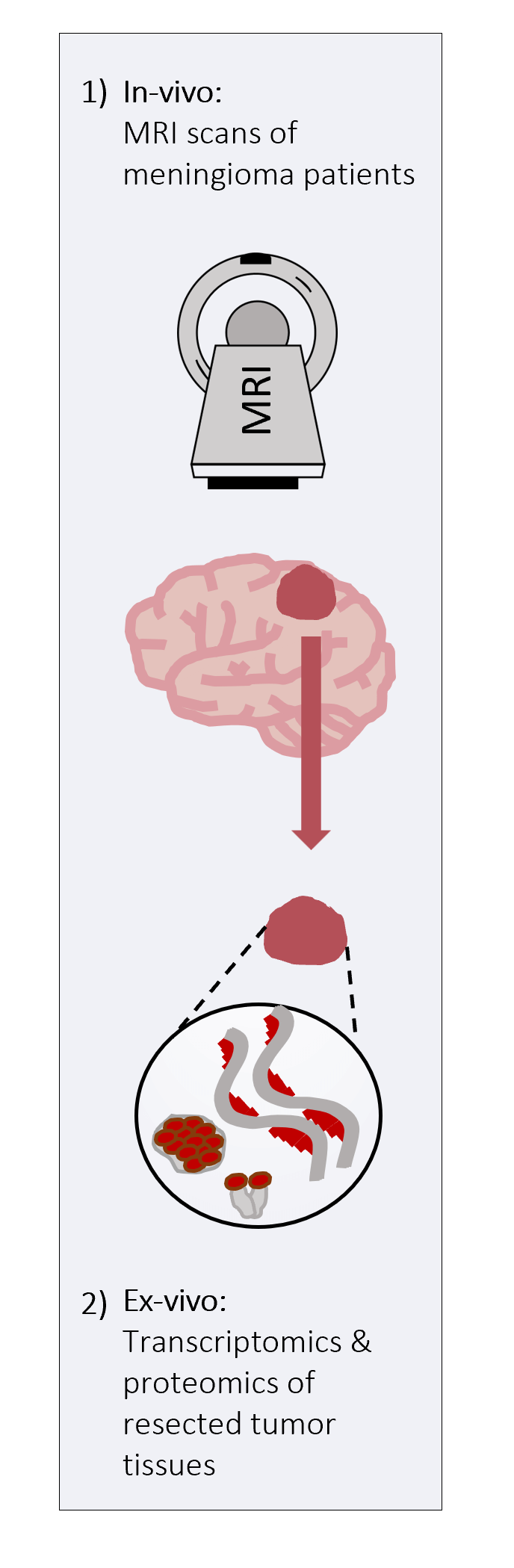
As part of a collaboration led by Dr. Tal Shahar, who currently directs the brain tumor unit at Sourasky Medical Center in Tel Aviv, and the doctors Dr. Nebo Margalit, director of the neurosurgery department, and Dr. Eliel Ben-David, director of the neuroradiology unit at Shaare Zedek Medical Center, the researchers video Because the method of measuring the MRI The new one correctly represents the iron homeostasis data in the specific tissue being measured. In an experiment at Shaare Zedek Hospital, the team of researchers screened subjects with active brain tumors of the meningioma type using the MRI the new one and predicted the iron data based on it. After removing the tumor in the neurosurgery department and taking a biopsy, the levels of the proteins relevant to the iron balance in the tumor tissue were tested in the molecular neuro-oncology laboratory. . To the joy of the researchers, a high agreement was found between the iron data shown by the MRI Before the surgery to measure the proteins from the actual tumor tissue.
In addition, the samples were also sent to the laboratory of Dr. Naomi Habib from the Lily Safra Neuroscience Center (ELSC) at the Hebrew University specializing in the characterization of genetic expression in brain tissue. It was found that there is a close relationship between the new measurements b MRI and the gene expression profile related to the iron economy that the tumor showed.
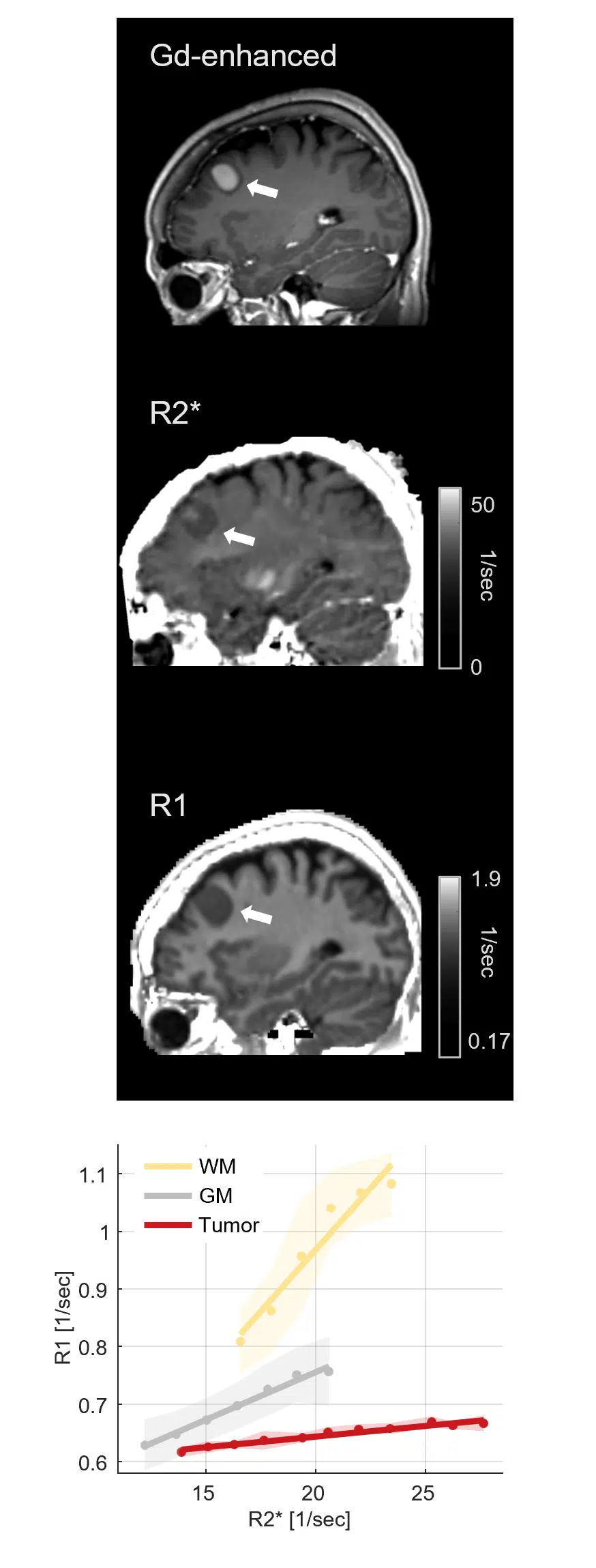
Even among those required for inspection MRI simple, showed the measurement of MRI The new one developed by the researchers has advantages. By making it possible for the first time to differentiate very well between tumor tissue and healthy brain tissue, even without the injection of a contrast agent which can be dangerous to the subject.
Today, in order to identify brain tumors, a contrast agent is injected before the scan MRI. Along with its main advantage, in that it makes it possible to distinguish between healthy tissue and tumor tissue, concerns have been raised in the past that it is not fully cleared from the brain and may cause damage to the subject. Use of the new measurement method may in the future allow the identification of tumors without the use of a contrast material that may be toxic to the patient and harm him.
More of the topic in Hayadan:

One response
It is generally accepted and recommended to give an explanation next to the graphs and pictures.