Scientists from the Technion and the Helmholtz Institute in Munich have developed a new technology for rapid XNUMXD imaging of extensive nerve cell populations at an unprecedented depth
Scientists from the Technion and the Helmholtz Institute in Munich have developed an innovative opto-acoustic technology for brain imaging, which enables three-dimensional monitoring of areas containing millions of nerve cells (neurons) with unprecedented speed and depth. This technology has enormous advantages over conventional optical imaging.
The research, which was published in the prestigious journal Light: Science & Applications from the Nature group, was carried out in collaboration between Prof. Shai Shom from the Faculty of Biomedical Engineering at the Technion, Technion graduate Prof. Daniel Rezensky from the Helmholtz Center and the Technical University of Munich, and a team of researchers that included Dr. Louis Dean- Ben and Dr. Gali Sela.
Comprehensive and deep imaging of the brain is a necessary condition for understanding how the brain works in health and disease, and observing nerve cell populations in the brain and their interactions is a central goal in the development of brain imaging technologies. Hence the importance of the new method, which allows, according to the researchers, "to observe a huge amount of nerve cells and see how the messages pass between them like wildfire. Our method makes it possible to observe the activation of large neural circuits, and even the entire brain in the case of a small animal. In our estimation, the technology will help in the development of new treatments for neurological and psychiatric disorders."
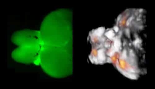
The new system, named FONT (functional opto-acoustic neuro-tomography), is based on short laser pulses that cause tiny and temporary oscillations of the tissue, resulting in the creation of ultrasound waves. These fluctuations are monitored by dedicated detectors and translated into three-dimensional images of the tissue. The innovative technology is based on the discovery of Prof. Rezensky and others, according to which the proteins sensitive to calcium concentration have an opto-acoustic signature that enables monitoring of brain activity. This monitoring is based on rapid changes in calcium ion concentrations accompanying brain activity.
The researchers tested the new system on the brains of adult zebrafish. However, they point out, the new technology enables the imaging of much larger brains with a volume of up to hundreds of cubic millimeters - up to 1,000 times the maximum volume possible with the existing methods of optical microscopic imaging, which are very limited in their depth of penetration. The state-of-the-art system also has a huge advantage in the time dimension - it performs the imaging at a rate of 100 volumetric images per second and thus enables the monitoring of fast processes such as electrical activity in neurons.
For the full article:http://dx.doi.org/10.1038/lsa.2016.201
