Technology developed by researchers at MIT and the Technion will improve the efficiency of X-ray machines and CT systems and their speed of operation. Prof. Ado Kaminer, head of the AdQuanta laboratory, participated in the study published in Science

A technology developed by researchers at MIT and the Technion is expected to lead to a leap forward in the efficiency of X-ray machines and CT systems and in the speed of their operation. Prof. Ado Kaminer, head of the AdQuanta laboratory, participated in the study published in Science For Robert and Ruth Magid.
The discovery of X-rays in 1895 brought about a dramatic change in the world of medicine. William Roentgen's invention led to the use of X-rays, also called X-rays, in mapping the internal tissues of the human body. Although the X-ray is mainly used to map the condition of teeth and bones, today it is used for other purposes, including the detection of muscle tears and mammography of the breast, as well as non-medical uses such as testing the quality of various industrial components.
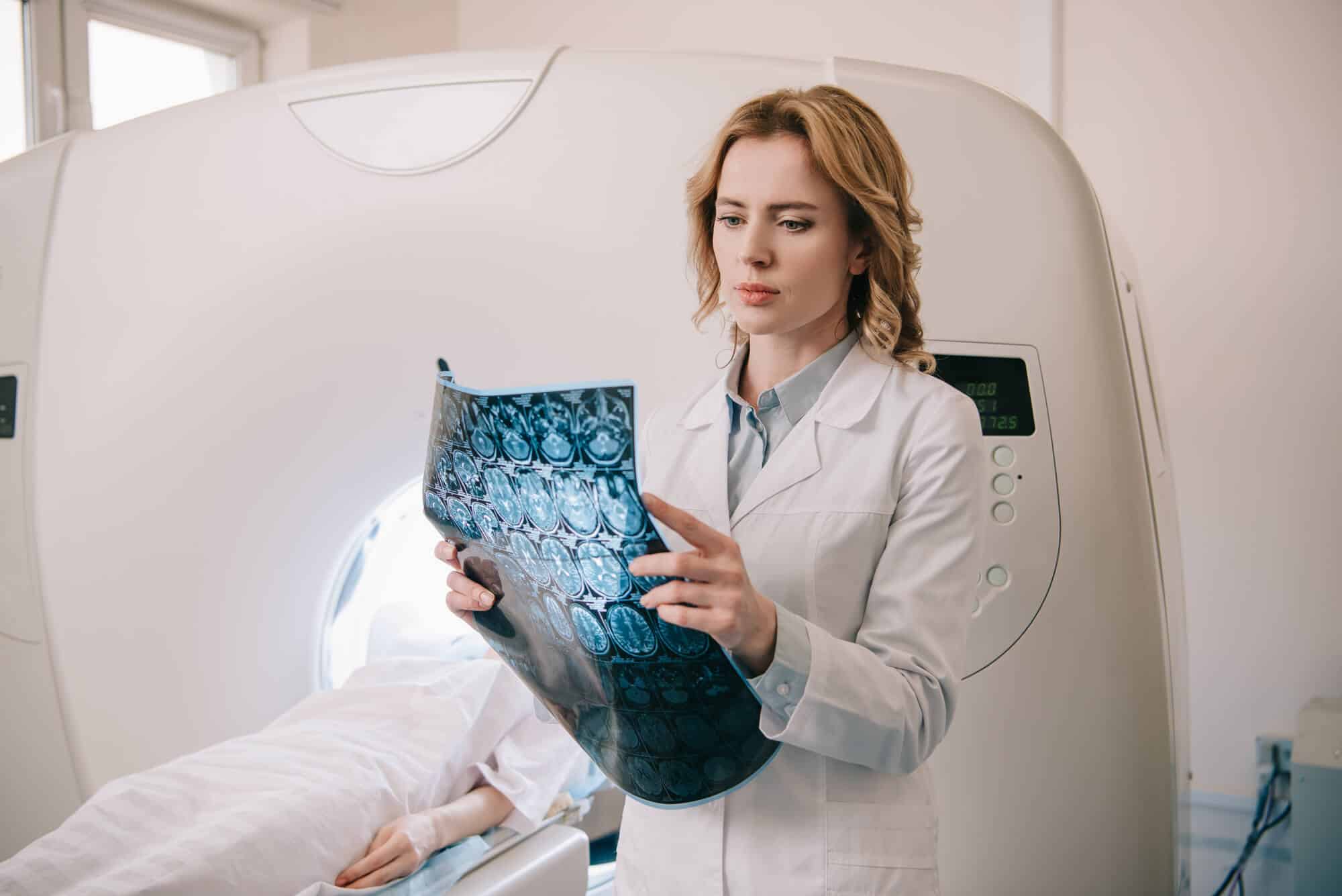
CT technology, which was only invented in 1972 - almost a century after the first X-ray machine - makes more sophisticated use of the same radiation; By editing many photographs from different angles, and their computer analysis, it provides a complex XNUMXD image of the scanned area. In the medical applications of X-ray and CT, X-rays are launched into the subject's body and are picked up on the other side by detectors called scintillators ("sparkles" in Hebrew). These detectors pick up the high-energy photons of the X-rays and convert them into "normal" photons - light radiation in the visible spectrum. This radiation hits a photographic plate and this is how the clinical image of the inside of the body is obtained. A similar process is also used in scientific research (in an electron microscope) and in optics (night vision systems, for example). One of the bottlenecks in the scintillation process is The minority of photons that emit scintillators as a result of their dispersion to all angles (called isotropic emission). Due to this phenomenon, a significant part of these photons do not reach the photographic plate and the result is a relatively low resolution of the resulting image. In principle, this can be covered up by significantly increasing the x-ray radiation, but it is customary to avoid this for obvious reasons - strong radiation is dangerous for human cells, and it is especially dangerous for children and people who undergo these scans as a matter of routine.
So far, the main effort in improving scintillators has focused on trying to build scintillators from new materials, a challenge in which progress is few and far between. The research group from MIT and the Technion took a different approach: instead of developing new scintillation materials, they changed the surface of the existing materials in a way that improves their efficiency. The researchers estimate in the article that it is a 10-fold improvement in this resolution, but it is possible that this is an underestimate and the actual improvement is closer to 100-fold.
The inspiration for the aforementioned technological improvement idea came from Yaniv Korman, a doctoral student in the Viterbi Faculty of Electrical and Computer Engineering who is conducting his doctoral research under the guidance of Prof. Ado Kaminer. In an article published in the summer of 2020 inPhysical Review Letters (See additional coverage atPhysics Today) The two presented a new approach in the detection of X-ray radiation - by improving scintillators from existing materials by processing the materials on a nanometer scale, which is specially adapted to the wavelength of the scintillation light emitted from them. In the same article, the two emphasized that an applied presentation of the system may take a long time; In retrospect it now turns out that in less than two years the first system was built in collaboration with MIT researchers and under their leadership.
Although more follow-up research and applied effort will be required to integrate the improved scintillators into actual medical systems, Prof. Kaminer believes that the method he developed with his colleagues "will lead to a dramatic improvement in the quality of the image obtained while significantly reducing the patient's exposure to radiation - two central goals in the development of new X-ray-based diagnostic technologies . The new method will also improve non-medical technologies based on the creation and detection of X-ray radiation."
Two world-renowned scientists, who did not participate in the said study, enthusiastically responded to the publication. "The paper presents a very significant achievement," said Prof. Rajiv Gupta, a neuroradiologist at MGH (Massachusetts General Hospital) and a professor at Harvard Medical School. "It demonstrates the fact that the technology improves X-ray scans 10 times, and even if it improved them 'only' twice, it would be a real revolution in this field." According to Prof. Eli Jablonovich, an electrical and computer engineer at the University of California, Berkeley, "Although the application of the approach in medical applications still needs to be demonstrated, this is a very original and high-quality work on a subject that has not received the attention it deserves: scintillators based on photonic crystals."
MIT researchers Prof. Marin Suliacic, Nicolas Rivera, and Charles Rock-Carm are partners in the study.
More of the topic in Hayadan:
