This is how the retina separates the colors and increases the sensitivity of the eye 10 times
An interdisciplinary research group from the Technion revealed for the first time, in the journal Nature Communications, the important optical role of the retina in improving light absorption in the eye. This study proves that the retina is a sophisticated optical organ, and not just a system for converting light into electrical signals and for the primary processing of visual information.
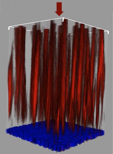
The research shows that the light, which enters the eye through the pupil and falls on the retina, is separated according to its color by the glial cells (support cells), which function as optical fibers within the retina. These cells, it turns out, efficiently collect and conduct the light in the green-orange range to the cones responsible for day vision, and allow the light in the blue-violet range to disperse to the cones responsible for night vision. This separation of colors allows up to a 10-fold improvement in peripheral daytime vision, without harming night vision. This is a sophisticated evolutionary solution that matches the structure of the retina, where the light receptors are located behind the layers of nerve cells and nerve extensions that process the image.
In the study, a computational model was built that predicts the phenomenon in the peripheral retina of humans and other mammals that are active during the day. This model was verified in laboratory experiments, in which the light moving in the glial cells was measured in the depth of the retina up to the light receptors. In these measurements it is checked - for each color in the visible light range - how much light passes inside the glial cells and how much light is scattered outside the cells. As mentioned, the spectral distribution was found to be suitable for the computational model, and the light indeed moved along the glial cells until they reached the receptors.
The research was conducted in collaboration between the faculties of physics and medicine at the Technion, by the research students Amichai and Vasadi Spuri and their supervisors, professors Edo Perlman and Erez Ribak.

One response
Wow!!! It's all a blunder???