So far, they have succeeded in transplanting engineered muscle tissue with only small blood vessels

The Technion researchers for the first time implanted engineered tissue with main blood vessels in an abdominal muscle with a severe injury. This is how the prestigious scientific journal PNAS publishes. Previously, researchers were able to transplant engineered muscle tissue with small blood vessels. The breakthrough may save complex surgeries in the future and the Technion registered a patent for it.
Professor Shulamit Levenberg from the Faculty of Biomedical Engineering and the Russell Berry Institute for Nanotechnology Research, explains that in the current study, the researchers created engineered muscle tissue with a major vascular system (FLAP), to repair large defects. The Technion researchers were able to reproduce a large defect in the abdominal wall of a mouse.
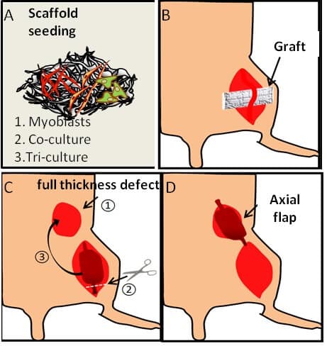
In the field of reconstructions, tissues can be used in two ways: 1. GRAFT, where the researchers take a tissue and transplant it. The blood vessels in the new environment enter the tissue and nourish it.
2. FLAP (hanger) - transfer tissue to the defect area without cutting off the blood supply to it. This stage is used when the target environment is damaged, the damaged area cannot provide support with new blood vessels and a tissue with a large blood supply is needed, such as in cases of cavity closure, or exposed bone, cartilage or tendons.
In tissue engineering, replacement tissues are developed for transplantation. The engineered tissue in this study is a three-dimensional tissue produced on a biodegradable, porous polymer skeleton, on which are endothelial cells, support cells (fibroblasts) and muscle cells (myoblasts). The Technion researchers grew the tissue in the laboratory and then implanted it around the artery and veins in the thigh area. She was then transferred to a stage called FLAP, where tissue with its main blood vessels is taken and connected to the blood vessels in the environment where it was implanted, to close a large defect in the abdominal wall.
The GRAFT stage is good for areas where the damage is relatively light. But when the damage is severe, it is not enough and therefore the importance of the current breakthrough is great.
The research was carried out in collaboration with Dr. Yulia Shandlov (who was a doctoral student of Professor Levenberg at the time of the research) and Dr. Dana Aguzi, from the plastic surgery department at the Rambam Medical Center, who was recently appointed director of the plastic department at Kaplan Hospital.
The research is part of a project by Professor Shulamit Levenberg, funded by the European Union (ERC) in the amount of one and a half million euros.

One response
I thought they were implanted in a human. For now, just a mouse.