The applicability of the new technology is demonstrated in the contexts of local cell transplantation, drug transport for controlled local release over time and 3D bioprinting. The mechanical properties of the scaffolds can be adjusted according to the target tissue and the desired drug release rate
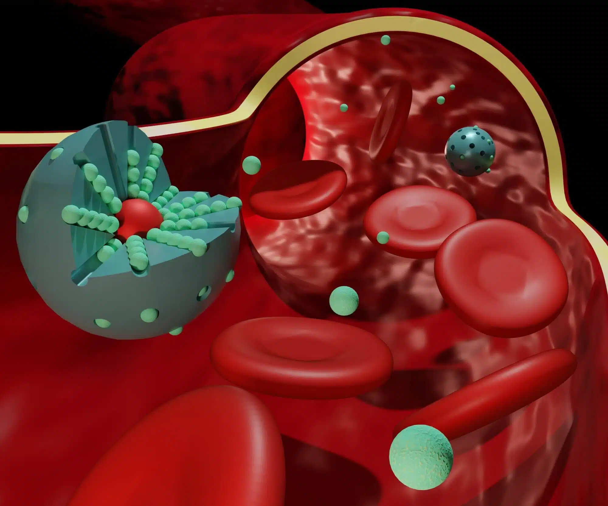
Newspaper Small Methods Presents a breakthrough in drug delivery and tissue transplantation with the help of ultrasound - a development by the researchers of the Faculty of Biomedical Engineering at the Technion. Prof. Shulamit Levenberg's research group, led by post-doctoral student Lior Debi and doctoral student Majed Mash'or, developed an innovative method for bioprinting living cells and tissues deep in the body in a non-invasive manner by soaking sound waves outside the patient. The technology is based on liquid materials that are sensitive to sound (acousto-sensitive materials) that solidify quickly under the influence of ultrasound from an external source. This process does not require invasive exposure of the treated tissue, radiation or other harmful actions.
The applicability of the new technology is demonstrated in the contexts of local cell transplantation, drug transport for controlled local release over time and 3D bioprinting. The mechanical properties of the scaffolds can be adjusted according to the target tissue and the desired drug release rate.
In the field of tissue engineering, which aims to produce functional tissues for transplantation for the purpose of restoring dysfunctional tissues and organs, another approach has developed in recent years of building the engineered tissues directly in the patient's body using various printing methods. Direct printing of the tissues built from living cells and supporting scaffolds requires an invasive procedure that includes massive exposure of the treated site and carries risks for the development of complications, peripheral injuries and infections. In the innovative method developed at the Technion, cells or drugs are transported in liquid biological ink directly to the treated area deep in the body through direct injection or catheter. The engineered tissue is then printed using sound waves transmitted from an ultrasound transducer external to the patient. In this way, engineered tissue can be built deep in the body without exposing the treated site.
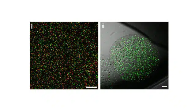
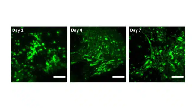
In a work published after peer review in the scientific magazine Small Methods The researchers presented several innovations. First, the team was able to demonstrate for the first time the construction of tissues with functional living cells such as heart cells, by means of the external soaking of sound waves. The cells continued to function and beat after the procedure. In addition, endothelial cells formed vascular networks and were alive and functional for at least a week after printing. Second, the researchers showed the possibility of loading the sound-sensitive bio-ink with anti-cancer drugs for a controlled and delayed release over time. In this way, prolonged local treatment can be carried out to reduce unwanted effects in healthy areas.
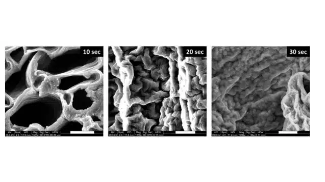
Third, the researchers showed that it is possible to control the mechanical properties of the printed scaffold by changing the absorption profile of the sound waves (duration and intensity) so that it optimally matches the target tissue being treated or to control the desired drug release rate.
The proof of feasibility in this study is a basis for further research for a variety of biomedical applications and opens a window to innovative therapeutic approaches in tissue engineering, controlled release of drugs and bioprinting.
For the article in Small Methods
More on the subject on the science website

One response
Lovely but unnecessary. It is known that body systems can be repaired using frequencies. With a frequency generator and diagnostic system each kit costs maybe NIS 3000. And you can use it without limitation and without the story about liquids that solidify and all kinds of stories (sounds to me like a story designed to make it sound like a complex and reliable treatment and also less accessible. To sell complex treatments at a more expensive price than "just" frequency treatment)